


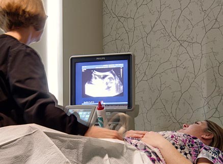
 Ultrasound imaging, also called sonography, is an amazing way of seeing the structure and function of the body using high frequency (but inaudible) sound waves. Sound waves are transmitted from special transducers or probes into the body part being examined.
These sound waves then bounce off and/or go through the organs of the body, returning to the transducer. The returning sound waves are converted into images by special computers and displayed on a screen instantly.
Being able to view the images instantly or in real time affords advantages in looking at body parts in any position needed and in assessing movement. Looking right where you hurt is easily done!
Special transducers can be used to obtain 3 dimensional images of different structures, most frequently of the female pelvic organs and fetuses. Certain structural changes are best shown when viewed in 3D, including things like uterine malformations or masses and fetal facial defects.
Doppler ultrasound is a special technique based on the principle of the Doppler effect, that is the change in frequency of sound with movement (think train horn!). This allows us to visualize and assess blood flow in blood vessels and solid tissues.
Assessing the fetus during pregnancy may be the most familiar ultrasound test, and it has indeed revolutionized the practice of obstetrics. Fewer maternal and fetal deaths can certainly be attributed to the use of ultrasounds during pregnancy, allowing us to quickly assess for abnormal fetal position, risk of bleeding from abnormal placental position and fetal anatomy.
Ultrasound has uses throughout the body, from looking at the neck vessels for narrowings in those at risk of stroke, evaluating breast masses, looking for gallstones, assessing liver blood flow in cirrhosis, evaluating the uterus and ovaries, finding the source of scrotal pain to assessing tendons and muscles for tears. Most body parts can be evaluated with the exception of the adult brain – the skull is too thick for the sound waves to penetrate – and the lungs- air reflects the soundwaves.
Ultrasound is a highly sophisticated, safe, and painless exam that can help in screening for diseases, like abdominal aortic aneurysm or breast cancer, or in diagnosing the source of your symptoms, like abdominal pain. Our specially trained staff will work to make sure you are comfortable and your questions are addressed.