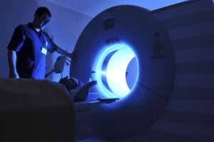
MRI / Open MRI
YOUR MRI AT DIAGNOSTIC IMAGING CENTERS
Magnetic Resonance Imaging, or MRI, is a noninvasive way of imaging the internal structures of the body using magnets, radiofrequency waves and powerful computers. MRI uses no x-rays or ionizing radiation. Images are created in thin slices through the body part being examined, and can be done in any orientation. Any body part from the head to the toes can be imaged.
MRI safety is our top priority! Because the powerful magnet is ALWAYS on, all removable metal such as jewelry, watches, hairpins and cellphones will be removed. These metal objects can become dangerous missles if allowed to enter the MRI suite. The magnet can also interfere with certain implanted devices as indicated below. Other metal within the body can create problems with the images. For these reasons, you will be carefully screened prior to your MRI both at the time of scheduling and prior to entering the room.
MRI Precautions
If you have any of the following, please discuss with our schedulers and technologists, as you may not be able to undergo MRI:
- pacemakers or implantable defibrillators (there are a few pacemakers which are MRI compatible, but special precautions must still occur with these; patients with other pacemakers cannot enter the MRI suite)
- cochlear (inner ear) implants
- neurostimulators
- some aneurysm clips
- implanted medication pumps
- catheters with metal tips
- some medication patches
If you have the following, MRI can usually be done, although special precautions may be necessary:
- metallic spinal rods
- joint replacements
- plates and/or screws for bone repair bone repair
OPEN VS.
TRADITIONAL MRI
If you are above average in size or have severe claustrophobia, an open MRI may be an option. The magnet is positioned above and below you, with the sides open. The open MRI uses a different strength magnet, and imaging on the open system takes longer. There are some exams that cannot be performed on the open magnet (like breast and prostate MRI), so speak with our staff if you have questions.
IV CONTRAST
Some MRI studies may be done prior to, and following, an IV injection of a contrast material. For MRI, the contrast is a form of gadolinium, a heavy metal. Contrast may be used to help assess the blood vessels and the way certain tissues take up the contrast. Prior to giving the contrast, you will be screened for any kidney problems as there are rare complications that can occur in patients when kidney function is low.
What to Expect
You will change into a gown. During the MRI you will be positioned on a table which is moved into an open-ended tube. The process of creating the images is loud, so ear protection is provided. Special coils that are able to send and receive the pulses used to create the images may be positioned around the body part being examined. Imaging takes between 20-45 minutes depending on the area of the body studied and the clinical situation. Motion affects the quality of the images, so it is important to hold still. You will be in view of and in constant communication with the technologist throughout the exam.
TIPS FOR AN EXCELLENT MRI
No jewelry, watches or metal can enter the MRI suite.
Let us know when you schedule your MRI if you have any metal in your body from prior injury or surgery, any implanted surgical devices like stimulators or a pacemaker. MRI safety is our top priority!
Relax and communicate. If you have claustrophobia, there are things we can do to help you through the exam. Oral sedatives can help if you have severe claustrophobia. If an oral sedative is used, you will need a safe ride home and a little extra time for the sedative to take effect.


