Mammography
MAMMOGRAPHY AT DIAGNOSTIC IMAGING CENTERS
MAMMOGRAPHY
Breast cancer remains a significant problem with 1 in 8 women facing the disease in their lifetime and with an estimated 40,000 women dying from the disease in 2016 alone. Screening mammograms have been proven to reduce the risk of dying from breast cancer. Starting screening at the age of 40 and screening every year saves the most lives, and so we recommend following those guidelines for most average risk (meaning no high risk factors like breast cancer in your immediate family) women.
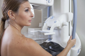
A mammogram is an image of the breast tissue obtained using low-dose x-ray technology, allowing us to see what is too small to be felt. Compression is used to eliminate motion which causes blurring and to spread out the normal breast tissue. Compression is used for both 2D and 3D studies for those important reasons.
In 2D mammography, two views of each breast, or 4 if you have breast implants, make up a routine mammogram.
In 3D mammography, views of each breast are taken by standing still as the gantry arm of the machine sweeps around the breast in an arc. Typically, two views of each breast are obtained in the 3D study.
Additional imaging, such as breast ultrasound or breast MRI may be needed about 10% of the time (less frequently if the initial screening is a 3D mammogram) — at our clinics we can do this additional imaging immediately; if there are areas of concern from a mammogram done on the mobile mammography coach, we will call and schedule follow-ups as soon as possible.
Have questions? Ask away! Our technologists are experienced and caring – and welcome your questions.
3D MAMMOGRAPHY
3D mammography, also called tomosynthesis is like traditional mammography in that it uses breast compression and low-dose x-ray technology. Images are obtained by the machine moving with an arc motion around the breast to create thin-sectional images of the breast tissue. This improves the radiologist’s ability to separate normal from abnormal tissue. This means we can find breast cancers at earlier and more treatable stages. This can also mean fewer additional work up mammographic views are necessary. We proudly uses 3D mammography equipment with the lowest radiation dose possible, matching the dose of our traditional 2D mammograms.
All women can benefit from this latest in technology, and an increasing number of insurers are covering the additional flat fee upcharge of $55 at time of service.
The following groups of women will likely benefit the most from 3D mammography: women with dense breast tissue, with a personal or family history of breast cancer, and those with breast cysts, masses or abnormal mammograms in the past.
SCREENING VS. DIAGNOSTIC
SCREENING MAMMOGRAMS
Screening mammograms are annual exams done for women with no symptoms. The aim is to screen all eligible women for signs of breast cancer. We recommend following the Society for Breast Imaging and American College of Radiology guidelines: yearly mammograms starting at age 40, if of average risk. No doctor’s order is required. These are covered without cost to you by insurance.
DIAGNOSTIC MAMMOGRAMS
Diagnostic mammograms are done for women with breast symptoms (pain, lump, breast discharge, etc.) or in women with breast cancer found in the last 5 years. A doctor’s order is required, and insurance coverage varies. At times, additional imaging such as ultrasound or extra mammographic views may be required. At our clinics, these can be done the same day, allowing you to leave with results in hand, just like our screening patients.
What to Expect
For the procedure, you will need to disrobe from the waist up. Deodorant will need to be removed- the metal in many deodorants shows up on the mammogram, mimicking one of the early signs of breast cancer.
After the mammogram, you will receive your results before you leave our clinic, or they will be mailed to you promptly if the screening was done on our mobile coach. Your report will also include a statement on your breast density.
Urgent results will be phoned in immediately to your referring physician.
For any breast imaging, having all of your previous images available is very helpful for accuracy. If your previous imaging has been done elsewhere, we will want to get them for comparison. Obtaining prior images from outside of Diagnostic Imaging Centers can be done by submitting a signed Breast Imaging Medical Records Release Form to our medical records team.
