More Mammography Views Taken? Good Reasons

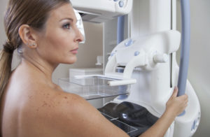
In a standard mammogram, you are probably familiar with the usual 4 views that will be taken: a side-to-side (MLO, we call them) and an up-down (CC) image of each breast. However, it is not unusual for additional views to be needed. We thought we’d review the most common reasons for additional views, so you don’t get anxious when more images are needed.
The mammography technologist or radiologist may find technical things on the 4 views, requiring additional images. These include the following:
Technical reasons:
-
Motion: If there is motion during the image capture, even sometimes just from breathing, it can cause blurring of the mammogram which makes the images difficult for the radiologist to read. Small calcifications in particular may be lost in the image if there is motion.
-
Not enough tissue: In order to find abnormalities in your breast, the tissue has to be included in the image. Breast tissue towards the chest wall muscles and tissue on the sides may be hard to get included in the image in some women. Extra views may be needed.
-
Folds: If the breast skin or breast tissue becomes wrinkled or folded during the mammogram, the folds may hide normal tissue.
-
Prominent tissue in axillary tail: The part of the breast that goes towards the armpit is important too! This is present in varying amounts from woman to woman and may require additional images.
-
Not enough compression on tissue: Compression is key to finding breast cancers in your normal tissue. If compression is not adequate, extra views may be done. If you have large breasts, compression may have to be done in segments in order to get good views of all your tissue.
For women with implants, remember 4 extra views with the implant pushed out of the way are also a part of the routine, standard mammogram.
There may be findings on the images or on physical exam that prompt additional diagnostic views. This is seen in the following scenarios:
Diagnostic reasons:
-
A lump or change you feel in your breast: If an abnormality is detected either by your self-exam or by your clinician’s annual breast exam, it sometimes requires getting additional views. The radiologist may request spot views of the area that is being felt to help define that area from the surrounding tissue.
-
Something the radiologist sees: Diagnostic views may be needed after a finding is discovered on a screening mammogram. This may just be an area that looks changed from your previous studies. It could be a lump which needs a spot view. Magnification views are done to help look at tiny calcifications in the breast.
-
Patients with breast cancer: Patients who have been treated for breast cancer may also need additional views and get diagnostic mammograms for the first 5 years after they are diagnosed.
So, remember that while 4 images is the standard, sometimes you will get a little extra attention. Whatever the reason, the additional views are done with care, to make sure the radiologist and ultimately you get the best information possible.
Image credit: Polanda by M Salhab et al via Wikimedia Commons. Licensed under the Creative Commons Attribution 2.0 Generic.
Originally posted 5/8/13 on mammographykc.com.
