What Is Fluoroscopy?
 Fluoroscopy is a way of imaging the body using x-rays that allows a radiologist to view the body in motion. A special machine uses low-dose x-rays that are sent through the body while the radiologist is observing the area of interest. Images project on a screen in the room.
Most commonly, this technology is used for imaging the gastrointestinal tract, the genitourinary tract, joints, and for guiding interventional procedures of many types.
For studies using fluoroscopy, you will be placed on a table. The table can be positioned in upright and horizontal positions, depending on what part of the body is being examined. For gastrointestinal studies, the table may be moved during the procedure, and you will be asked to change positions as well so that all of the area of interest is seen optimally.
For many procedures using fluoroscopy, some type of contrast material will be used to let us see the area of interest – including barium for the gastrointestinal tract and iodine for the genitourinary system among others.
In addition to the images on the screen in the room, additional images will be recorded, again using x-rays to give better detail of the areas of interest. These are similar to other x-rays obtained of the body. Fluoroscopy is an amazing way of seeing the body and its parts in real-time. We will be further discussing this technique in viewing the gastrointestinal system in upcoming posts.
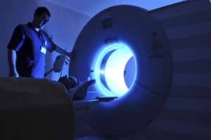
Image credit: Fluoroscope by Zereshk via Wikimedia Commons Copyright Public Domain
Originally published 7/14/14 on diagnosticimagingcenterskc.com.


