Shoulders, Knees and Toes, Knees and Toes
Over the next few posts we’re going to highlight some common injuries affecting familiar joints. As the title suggests, we’re starting near the top.
But before we get ahead of ourselves and start talking about these individual parts, let’s talk about how we image them…
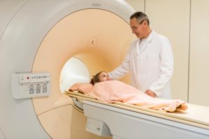
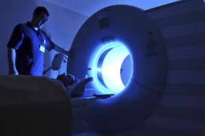
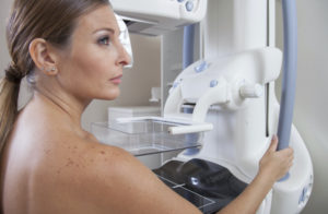
There are many techniques for imaging the body, and the ones we use depend upon the type of injury and the most likely tissues injured. Here’s a gimme: broken bones? We’ll start with an x-ray – perhaps a CT if it’s complex. Here’s a not-so-gimme: soft tissue injury like torn ligament? Options here include MRI, ultrasound and arthrograms.
First off, not every injury is imaged. Why? Sometimes a careful exam by your doctor can answer the question – imaging in these cases is not done, unless symptoms do not improve in the expected manner. There are carefully developed rules helping your doctors determine who will benefit the most from imaging in the case of many of the common injuries, for instance ankle sprains.
After your doctor’s initial evaluation, you may be sent for imaging. In many cases this will start with conventional films (x-rays) to exclude fractures or other bony changes. Beyond that, a patient will be directed based on the clinical concern.
Imaging of patients who have multiple sites of injury from a fall or motor vehicle accident for instance may be done with CT. This allows quick evaluation of bones as well as some types of soft tissue injuries. Multiple structures can be evaluated at the same time with CT, such as looking for fractures in the lower back, while also assessing the abdomen for signs of damage to internal organs.
Soft tissue damage, such as torn cartilage or ligaments, will often not be apparent on a conventional film. Visualizing soft tissues can be done with ultrasound, MRI or an arthrogram (where contrast material or dye is introduced into the joint space). We may follow the arthrogram with imaging with MRI or CT.
So… we will start our journey of the joints with your shoulders! (See you Thursday!)
Image credit: Head, Shoulders, Knees, and Toes by james.swenson13 via Flickr Copyright Creative Commons Attribution-NonCommercial 2.0 Generic (CC BY-NC 2.0)
Originally published 4/15/14 on diagnosticimagingcenterskc.com.

