Knee Arthrograms: Your Questions Answered!
Your questions answered about… knee arthrograms!
What is it?
- An arthrogram is a radiology study of a joint where contrast (sometimes called “dye”) is put into the joint with images then taken of the joint.
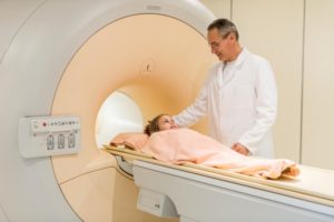
- The images can be taken with the fluoroscopy/x-ray system or with MRI or CT.
Why do we do it?
- The contrast distends (‘slightly expands from within’) the joint, allowing us to see soft tissue structures about the joint better.
- For the knee, an arthrogram may be requested by your doctor for the following reasons:
- Chronic knee pain
- Locking sensation
- To better assess meniscal tears
- To evaluate the knee after surgery
How do we do it?
- We will cleanse your skin at the knee with a solution to make sure we do not introduce infection.
- Local anesthetic or numbing medicine will usually be injected into the soft tissues to numb the knee – this part burns but the burning only lasts a short time, then you should just feel pressure.
- We will place a small needle into the joint, using our fluoroscopy machine and low dose x-rays to make sure we get the needle precisely in the joint space.
- The contrast material is then injected to distend the knee joint – iodinated contrast material if doing a conventional arthrogram or CT or dilute gadolinium, a heavy metal contrast material if being followed by MRI. This will make the knee feel tight.
Do I need to do anything to prepare for the test?
- No preparation is needed.
- We will ask for a list of your medications and drug allergies. If you have had a prior reaction to a contrast material, we will discuss the reaction with you and may adjust how we do the procedure.
Are there risks?
- The main risks from the procedure are bleeding and infection. If you are on blood thinners or aspirin, we will take care to hold pressure longer to prevent bleeding.
- The contrast material can sometimes cause reactions, but because the contrast material is going into the joint and not your blood vessels, the risk of reaction is very low.
Anything to know after the procedure?
- The contrast material sometimes irritates the joint causing pain. We recommend applying ice bag to the knee for about 15 minutes 3 or 4 hours after the study is done.
- There are no restrictions following the procedure.
- The tight sensation will wear off as the body resorbs the contrast over the next 24 hours. Moving the knee will help.
Image below: Yellow highlights the area behind the kneecap that has been injected with contrast material for the purposes of this MRI.

