MRI: It’s a Magnet!
 Radiology can be confusing. How these exams work can confuse the smartest people. As radiologists, we get lots of questions. Which exams use radiation and which do not? How safe is the exam?
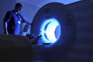
Today we will attempt to answer some of your questions and concerns about magnetic resonance imaging or MRI. MRI – how do we make images? MRI or magnetic resonance imaging, is one way of viewing the body that uses NO radiation (a plus!). We can create amazing images of all parts of the body with… a magnet! MRI machines are loud, clunky-sounding machines made up of a giant magnet. The noise comes from the second part of the name – resonance, or radiofrequency waves. This combined with computers creates images of the body. And, oh, the things we can learn about you with this technology.
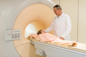
The MRI Experience Having an MRI involves being positioned on a table and moved inside the MRI unit. The inside of the MRI is called the bore and is basically a long tube the size of which varies. We center the body part being evaluated within the bore.
Bore sizes and configurations vary depending on the magnet strength and configuration of the MRI unit.
“Open MRI” units have a more open environment for imaging. They are often used for the claustrophobic patient or larger patients. The open MRI units can have limitations of longer exam times and lower quality of images. This is because the signal created and used to make the images is directly related to the magnet strength – which is lower for some open magnets.
High field magnets or traditional MRI trump an open magnet when we want imaging speed and precision, so it’s highly encouraged when at all possible. It can be done! There are many ways we can help our patients be comfortable while getting the highest possible quality images.
Over the next few days we’ll talk more about MRI safety as well as limiting patient discomfort in the machine.
In the mean time, cheers to your best possible health!
Image credit: Magnet by AJ Cann via Flickr Copyright Creative Commons Attribution-ShareAlike 2.0 Generic (CC BY-SA 2.0)
Originally published 10/6/14 on diagnosticimagingcenterskc.com.


