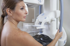
Mammography Saves Lives (in Just a Hundred Years)
 Science can move pretty quickly – these days. But medical research is always going to take time. Doctors and research-scientists want to be careful – extremely careful – about how we approach changes and advancements in care of patients. We want to be highly effective, quick, preventative, and oh yes, there’s that oath about “do no harm” – so we want to keep discomfort to a minimum. These are our goals when taking care of YOU!
The history of mammography is full of colorful characters, we promise.
Mammography isn’t a perfect science (people still suffer afflictions) but it is worlds better than where we started. Delving into breast cancer started with physically delving in – surgeries and biopsies. Then x-ray technology offered a non-invasive way of imaging the body. Then x-rays used less radiation. Then they became targeted at certain body parts – like with mammograms – and became even safer. Then… mammograms became digital and reduced the exposure rate while increasing the precision. And all this took a hundred years. There are many people to be grateful for progressing breast health, and we’re only able to name a few, lest this blog post read like a genealogy table. But we’ll toss a few important names out here.
Radiology (or roentgenography) wasn’t discovered until the late 19th century. It took another dozen plus years for it to be applied specifically to the breast, by a caring German physician named Dr. Albert Saloman. Saloman performed over 3,000 mastectomies in his lifetime, and owing to those experiences, he sought better ways of solving the breast cancer problem. Instead of surgeries and biopsies, he looked toward imaging. He originated using roentgenography on the breast specifically. His tireless work was interrupted by WWII, when he was forced to serve in a concentration camp before his escape to the Netherlands where he continued his work for the rest of his life.
After all of Dr. Saloman’s research, mammography was just around the corner.
That corner took a little longer to turn. It wasn’t until the 1920s-30s when research was going on simultaneously in the U.S., Europe, and South America that further progress was made. In the 1930s another German scientist, named Walter Vogel, published a paper confirming that cancerous and noncancerous cells could be differentiated in an x-ray of the breast.
In the U.S., Dr. Jacob Gershon-Cohen wrote passionately about the history of mammography, with deep respect for his colleagues. He published a paper that we couldn’t peel our radiologist-eyes off of. (And we have good faith that you, dear reader, have the capacity to understand it. You’re smart enough to be reading this, after all.) Dr. Gershon-Cohen is known for advocating what we today call screening mammograms – regular check-ins to confirm the absence of breast cancer, or to catch it in the earliest stages possible. Dr. Stafford Warren developed a stereoscopic technique for more detailed views of the breast, though it was expensive and somewhat complicated. He abandoned his work in mammography when called to serve the US government work on other things, such as the Manhattan Project.
In France, Ch. Gros developed the senographe, a technique for reading soft tissue (breast as opposed to bone) with x-rays for more accurate readings. In the U.S., Dr. Robert Egan found he could predict with near precision cases of breast cancer in one three-year study (discovering 238 out of 240 cases in one thousand women).
Finally in 1986, Patrick Panetta and Jack Wennet procured the patent for compression mammograms, the procedure that is commonly used now.
It is thanks to this progress that millions of women have been screened and millions of lives saved. And all it took was a century.
Originally published 7/2/13 on mammographykc.com.
