Imaging After Breast Cancer
 Our mantra is: yearly screening mammograms for all women over 40 with breasts. We have not discussed in any detail times when imaging is required after a breast cancer diagnosis or after surgery. There are a few special considerations to take into account and a few different things to expect when you have had prior breast surgeries.
-
If you’ve had a lumpectomy…
Typically, a six month mammogram is scheduled for the side of the surgery. This helps to establish your new baseline appearance of your breast tissue on the mammogram. There will be scarring at the surgical site, but the amount of scarring is quite variable.
If there a suggestion of fluid at the site of the surgery (not uncommon!), sometimes an ultrasound may be needed. Depending on your type of cancer or on the findings on your mammogram, your doctor may take a cautious route and request follow-up mammograms every six months for up to three years on the side that had the cancer. Yearly studies of the other breast are usually sufficient.
-
If you’ve had radiation therapy…
As with a lumpectomy, 6 month mammography on the affected side may be requested. This establishes your new baseline. You will have scarring and often thickening of the skin in the area of radiation. This is normal and variable.
We typically like to see the scarring become less prominent in the years following your radiation. If at any time the scarring appears more pronounced, further imaging, often with breast ultrasound and or breast MRI may be necessary to help separate normal scar from possible recurrence.
-
If you’ve had a mastectomy…
If you’ve had a mastectomy, the “surgical bed” or chest wall on the side of surgery will be followed with clinical exams. If there is a change on the clinical exam, like a focal bump, mass or thickening, imaging may be required.
This most often starts with ultrasound, although occasionally mammography may be requested. MRI of the breast may be needed based on the other imaging results or for difficult cases. If you have had breast reconstruction using a flap type of surgery (where muscles and tissue are moved from one part of the body to recreate a breast), the decision to image is based on the preferences of both the surgeon and/or patient. The same is true for breasts reconstructed with implants after mastectomy. Most often, these patients do not require routine imaging on that side. Imaging may be necessary if there is a change on your physical exam.
-
If you have a personal history of breast cancer…
Remember, you have a significant personal risk factor for breast cancer in any remaining breast tissue after treatment. The need for screening will continue for the rest of your life.
-
If you have had a breast biopsy…
Six month mammogram on the side of biopsy may be requested, and again helps to establish the new baseline appearance of your breast tissue. The amount of scarring is usually minimal from needle biopsies, but will vary from person to person and depending on what exactly was done. The location of the biopsy is noted with a metallic marker so that the site is known precisely.
-
If you have had breast reduction or breast implants…
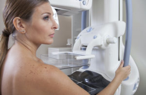
Routine yearly screening mammography is recommended for women with either breast reduction or augmentation. For implant patients, additional views will be obtained with the implant moved out of the way for optimal compression of the breast tissue. This means 8 images for a standard implant study versus the usual 4.
For reduction patients, no special views are required. There may be areas of scarring and often benign calcifications in the breast tissue or skin following the surgery. Sometimes these will require special views, such as magnification views.
Some women are concerned that mammography after surgery may be more painful. For the majority of patients this is not the case. Rest assured that your mammography technologist will work with you if you are experiencing tenderness or pain. Communication is key!
Originally posted 1/31/14 on mammographykc.com.
