Headaches and Head Issues #2: Looking Inside Your Head
 So… there will be times when a headache prompts further evaluation. Imaging can be used to study the brain and its surrounding tissues. CT and MRI are both common imaging techniques for evaluating the brain and adjacent tissues when imaging for headaches is indicated.
For sudden onset headache, thunderclap headache, and headache following trauma in the past 48 hours, we often start with CT of the head.
CT Scans The initial CT imaging is done without contrast; images are obtained through the skull while the patient lies still. This takes only a few minutes. From this we can see hemorrhages in and around the brain – one of the serious causes for headaches that can be seen from both traumatic and non-traumatic causes.
Occasionally, the noncontrast study will be followed by postcontrast imaging after an IV injection of iodine-containing contrast – this highlights the vessels and demonstrates abnormal enhancement in the brain, such as masses.
CT uses radiation to make its images – therefore, use in pregnant patients will generally be reserved for special indications and circumstances.
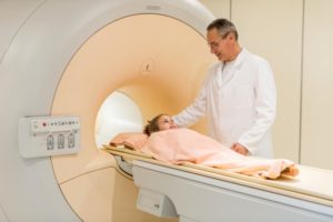
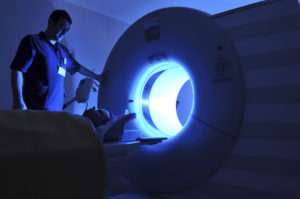
MRI Scans If there are any neurologic changes associated with your headaches (things like numbness, loss of strength or confusion) imaging with MRI may be requested. An MRI shows the internal structure of the brain in great detail. Masses and areas of abnormality from things such as strokes and multiple sclerosis are well shown with this modality. Because the procedure takes about 30 minutes to fully image the head, it does require the ability to lay on your back for a length of time. Images can be obtained both without and with IV contrast containing gadolinium, often times with both. Gadolinium contrast helps us look at vascular structure and for abnormal enhancement.
MRI can be used in some instances during pregnancy, but only after the first trimester is complete. No IV contrast is used for MRI in pregnancy.
Patients with pacemakers and other implanted surgical devices may not be able to undergo MR imaging. Let your doctor know of all surgeries and procedures prior to scheduling your MRI.
These exams can shed amazing light on the brain and its functions (or malfunctions). While we always work to image wisely, we can also image exquisitely. From the finest of endings to the largest of masses, we are able to have a noninvasive peak inside the inner workings of the brain. Through this we are able to get our patients on the road to their best possible health!
Image credit: Brain fMRI via Wikimedia Commons, Copyright Public Domain
Originally published 8/19/14 on diagnosticimagingcenterskc.com.


