Follow-Up Breast Imaging: What Might Be Next?
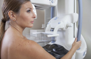
Sometimes, a 4-view mammogram will not provide all the answers about your breast health. Additional imaging at the time of your mammogram or even follow-up imaging may be needed. When it comes to these, there are many options which may seem confusing. A little enlightenment about possible additional imaging might help.
For breast imaging when patients are over the age of 30, most imaging will start with a mammogram, screening or diagnostic. If there is a finding on a screening mammogram that needs further work-up or if there is a breast symptom, we may ask for additional imaging as follows:
- Additional mammographic views: the standard 4 views may be supplemented by additional views in a different position, like straight from the side or with your breast tissue moved slightly. We may need to do views with compression of just one area of your breast, a “spot compression view” and for these we use a different sort of compression paddle. For some calcifications in particular we may need views obtained with magnification. All of these help problem solve findings on a standard mammogram. For breast implants, remember additional views with the implants moved out of the way are standard (known as “implant displaced” views).
- Breast ultrasound: we may request a breast ultrasound to give us more information about something we see on a mammogram, like a spot of tissue or an area of asymmetric tissue. Breast ultrasound can show soft tissues and areas of fluid like cysts well. Ultrasound is also done to look at an area you or your referring clinician might be feeling and possibly to fully evaluate areas of breast pain.
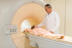
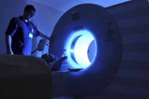
- Breast MRI: a breast MRI may be requested as a screening test for cancer in some women who have a high risk for breast cancer. We may also need MRI to help problem solve or for additional information about a finding seen with mammography and ultrasound. MRI shows anatomy of tissue as well as blood flow information, adding to what we can see with the other imaging techniques.
- Biopsies: a biopsy may be recommended after all of the imaging tests have been done and a tissue diagnosis is needed. Small amounts of tissue are taken from the breast and evaluated by a pathologist, a special doctor in a laboratory.
There are many options in breast imaging. In working towards a goal of finding breast cancer at its earliest stage when cure is possible, we may need to do more than a standard mammogram. If you need additional imaging, we hope this information will lessen any anxiety.
Originally posted 7/24/13 on mammographykc.com.
